Lower Body Bone Diagram - The lower limbs | Human Anatomy and Physiology Lab (BSB 141) - This area is commonly referred to as the calf.. Muscles and bones in human body 12 photos of the muscles and bones in human body how many muscles and bones are in the human body, how. There are two hip bones, one on the left side of the body and the other on the right. Folge deiner leidenschaft bei ebay! Legs are used for standing, and all forms of. The bones of the legs are those that make up the thigh, the lower half of the legs, and the feet.
The lower leg contains two major long bones, the tibia and the fibula, which are both very strong skeletal structures. The muscles of the lower back help stabilize, rotate, flex, and extend the spinal column, which is a bony tower of 24 vertebrae that gives the body structure and houses the spinal cord. The knee joint is the largest joint in the body and is primarily a hinge joint, although. Teachme anatomy part of the teachme series the medical information on this site is provided as an information resource only, and is not to be used or relied on for any diagnostic or treatment purposes. High resolution textures and displacement included.
Leg bone - Wikipedia from upload.wikimedia.org The knee joint is the largest joint in the body and is primarily a hinge joint, although. Female anatomy includes the external genitals, or the vulva, and the internal reproductive organs. The knee joint is the largest joint in the body and is primarily a hinge joint, although some sliding and rotation occur. Tibia is your leg's second biggest bone. Diagram femur bone diagram data pre. Your lower leg comprises of two main bones: Cheek bone (zygoma) upper jaw. The bones of the legs are those that make up the thigh, the lower half of the legs, and the feet.
The head of the femur forms the round ball of the 'ball and socket' joint of the hip.
The bones of the pelvis and lower back work together to support the body's weight, anchor the abdominal and hip muscles, and protect the delicate vital organs of the vertebral and abdominopelvic cavities. Anatomy of shoulder 12 photos of the anatomy of shoulder anatomy of nerves in shoulder, anatomy of posterior shoulder dislocation, anatomy of right shoulder, anatomy of shoulder labrum tear, anatomy of the shoulder games, human anatomy, anatomy of nerves in shoulder, anatomy of posterior shoulder dislocation, anatomy of right. Femur (2) tibia (2) fibula (2) patella (2) tarsals (14) metatarsals (10) phalanges (28) total number of bones=60. This is the longest bone in the human body, and is also known as the thigh bone. Tibia is your leg's second biggest bone. There are two hip bones, one on the left side of the body and the other on the right. Your leg bones are the longest and strongest bones in your body. Anatomy of the arteries and bones of the lower limb based on 3d pictures and angiogram (angiography). The lower leg extends from the knee to the ankle. This page is about leg bones diagram,contains aluminium plant safety: Leg bone diagram / arrangement of lower limb bones. Because of the important organs situated in the abdominal area, many health concerns stem. The lower leg contains two major long bones, the tibia and the fibula, which are both very strong skeletal structures.
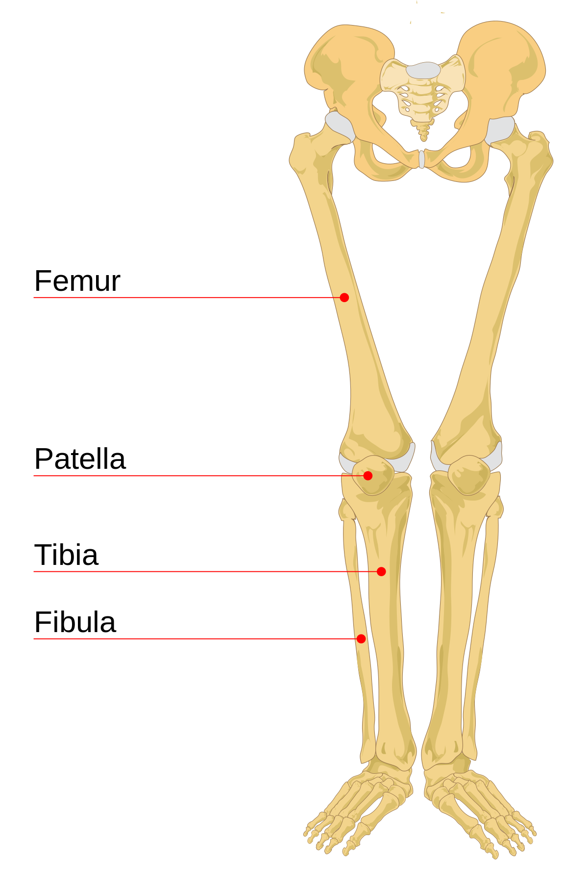
Other sesamoid bones can form in the joints of the hands and feet, but are not present in all people. The bones of the leg are the femur, tibia, fibula and patella. However, the posterior bony structure is different—lamina, pedicles and bony processes project off the back of the vertebral body. There is one bone in that region, which is known as the 'femur.' it is your body's largest bone. Lower leg muscle diagram blank.
Barcharts Muscular Origins & Insertions Quick Study Guide ... from cdn.teachersupplysource.com The knee joint is the largest joint in the body and is primarily a hinge joint, although some sliding and rotation occur. Reviewed by carol dersarkissian, md on may 18. The back supports the weight of the body, allowing for flexible movement while protecting vital organs and nerve structures. (right) the fibula and the tibia, bones of the lower leg. Human anatomy for muscle, reproductive, and skeleton. Female anatomy includes the external genitals, or the vulva, and the internal reproductive organs. Lower leg muscle diagram blank. From the front (or anterior), the vertebral body appears rounded.
Similarly, its base makes up a portion of your knee.
Posted in diagrams leg parts anatomy. Leg bone diagram / arrangement of lower limb bones. The arch surrounds the hollow vertebral foramen and connects the body to the bony processes on the posterior of the vertebra. Also called the shin bone, the tibia is the longer of the two bones in the. Femur (2) tibia (2) fibula (2) patella (2) tarsals (14) metatarsals (10) phalanges (28) total number of bones=60. Muscles and bones in human body 12 photos of the muscles and bones in human body how many muscles and bones are in the human body, how. Anatomy of the arteries and bones of the lower limb based on 3d pictures and angiogram (angiography). High resolution textures and displacement included. The lower leg extends from the knee to the ankle. Human anatomy for muscle, reproductive, and skeleton. Your lower leg comprises of two main bones: From the front (or anterior), the vertebral body appears rounded. Similarly, its base makes up a portion of your knee.
Your lower leg comprises of two main bones: This article looks at female body parts and their functions, and it provides an interactive diagram. Legs are used for standing, and all forms of. Folge deiner leidenschaft bei ebay! They hold up your body, and along with your muscles, keep you moving.
Skeletal System Diagrams | Anatomy bones, Anatomy and ... from i.pinimg.com Create your own flashcards or choose from millions created by other students. Diagram femur bone diagram data pre. Your lower leg comprises of two main bones: Teachme anatomy part of the teachme series the medical information on this site is provided as an information resource only, and is not to be used or relied on for any diagnostic or treatment purposes. The lumbar spine is the lower part of the back. The knee joint is the largest joint in the body and is primarily a hinge joint, although some sliding and rotation occur. Leg bone diagram / arrangement of lower limb bones. Health diagram skeleton human body anchor chart science bone pelvis human.
Bone diagram forehead (frontal bone) nose bones (nasals) cheek bone (zygoma) upper jaw (maxilla) lower jaw (mandible) breast bone (sternum) upper arm bone (humerus) lower arm bone (ulna) thigh bone (femur) collar bone (clavicle) toe bones (phalanges) ankle bones.
Leg muscles anatomy muscular system anatomy human muscle anatomy anatomy bones human anatomy and physiology body anatomy leg muscles diagram muscle diagram lower leg muscles. Understanding the anatomy of your lower spine can help you communicate more effectively with the medical professionals who treat your lower back pain. Bone diagram forehead (frontal bone) nose bones (nasals) cheek bone (zygoma) upper jaw (maxilla) lower jaw (mandible) breast bone (sternum) upper arm bone (humerus) lower arm bone (ulna) thigh bone (femur) collar bone (clavicle) toe bones (phalanges) ankle bones. The bones of the leg are the femur, tibia, fibula and patella. The bones of the legs are those that make up the thigh, the lower half of the legs, and the feet. Skeleton bones diagram 11 photos of the skeleton bones diagram anatomy bones diagram, axial skeleton bones, bones of the skeleton quiz, human body bones diagram, labeled diagram skeleton, skeleton diagram with bone names, skeleton system bones, skull bones diagram, human anatomy, anatomy bones diagram, axial skeleton bones, bones of the. Leg bone diagram / arrangement of lower limb bones. Also called the shin bone, the tibia is the longer of the two bones in the. Other sesamoid bones can form in the joints of the hands and feet, but are not present in all people. From the front (or anterior), the vertebral body appears rounded. Lower back pain us common. This page is about leg bones diagram,contains aluminium plant safety: Reviewed by carol dersarkissian, md on may 18.
Teachme anatomy part of the teachme series the medical information on this site is provided as an information resource only, and is not to be used or relied on for any diagnostic or treatment purposes lower body diagram. This is the longest bone in the human body, and is also known as the thigh bone.


0 Komentar